


2 trimester of pregnancy, ultrasound scan: decoding
Table of contents:
Many people are interested in what trimester of pregnancy is carried out biochemical analysis and ultrasound? 2 trimester of pregnancy do a comprehensive inspection, without exception, all women. It is necessary to determine the positive or negative answer regarding the data previously obtained, and when the symptoms appeared, in which are shown the ultrasound.
To Do an ultrasound (2nd trimester) only under certain rules of preparation for the procedure. Decoding of the received data is held in accordance with the same principles as in the first survey.
The Second screening when carrying a baby involves two stages:
- Conduct in the 2nd trimester of pregnancy ultrasound.
- Screening test - blood in the veins in the number of hormones.
Main indications for examination
The Research is conducted according to the same indications as in the first trimester:
- The Presence of bacterial or viral infection.
- Sickness, betrayed by inheritance.
- Diabetes.
- Diseases of the joints.
- Cancer of one of their parents.
- Pathology of chromosomal nature
- A history of miscarriage or spontaneous delivery.
- Having a baby with certain abnormalities.
- What Happened before the death of the fetus in the womb;
- Poor results of the first screening made on the period of 14 weeks and later showed abnormal development of the embryo.
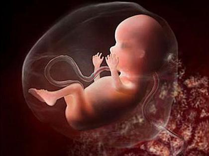
The rules of ultrasound 2 trimester of pregnancy makes it possible to ascertain the proportion of skeletal development of the child, to identify the condition of the ventricles of the brain, cerebellum, spinal cord and facial structures. In particular the attention on the nose, lips, eye sockets, the Atria of the heart, large vessels, kidneys, bladder and the digestive tract.
Recommended

How to choose a bike for kids?
If a toddler insists on not putting him in a wheelchair, then it's time to find another vehicle for him. Which one? A bicycle, of course. Kids will like this novelty, giving them a sense of independence. And parents, choosing it correctly, will not o...

Women's silver watches are an extraordinary accessory
A women's silver wristwatch is an exquisite and stylish piece of jewelry. Today, even a very demanding customer can choose the right model for herself.First, you need to sort out all the samples available for sale so that you can make the right choic...

Every girl should have a good epilator
Many girls who constantly take care of themselves are forced to use a razor. This often causes irritation, and unaesthetic hairs grow into the skin. It's not a pleasant feeling. But to stop removing hair on legs, armpits and in the bikini area is not...
Fotometria
Ultrasound Protocol 2 trimester of pregnancy involves marking:
- The location of the placenta and its thickness;
- Structure the degree of maturity of the placenta;
- The number of vessels in the umbilical cord;
- The amount of amniotic fluid;
- The condition of the cervix and uterine walls as well as its appendages;
- Indicators of fetometry.
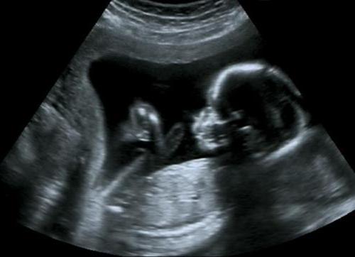
Fotometria is the measurement of the size of the fetus. It includes:
- Head size of the fetus, forehead and nape;
- Diameter head circumference and abdominal circumference;
- Cephalic index for estimation of the structure of the head;
- Length of tubular bones (hip, femur, large and small, humerus, ulna and radius) on both sides.
Decrypt ultrasound 2 trimester of pregnancy gives you the opportunity to establish its compliance with the development gestational age, assess fetal growth, confirm duration of pregnancy, identify the gap of development and the presence of pathologies.
Arrested development
Intrauterine growth retardation can be worn symmetrical and asymmetrical form. In the first case the development is proportional and ultrasound all indicators indicate the gap from the norm this pregnancy.
Asymmetric characteristics are as follows:
- Index abdominal circumference below the norm;
- Head size DBK and the norm for a long time;
- Increased the ratio of OG/of CSA and coolant/coolant.
Depending on the obtained results, we can distinguish three degrees of lag:
- A Lag of 2-3 weeks.
- A Lag of 3-4 weeks.
- For 5 weeks or more.
Assessment of the status of the internal organs of the fetus: 2 trimester of pregnancy, ultrasound
Cross-sectional head:
- Excludes abnormal structure in the form of lemon, banana and strawberries or pronounced brachycephaly, double contour of the head, which indicates dropsy of the brain;
- Examines the integrity of the bone structure of the skull.
Assessment of the brain:
- Ventriculomegaly shows enlarged ventricles of the brain;
- In the vascular system noted the presence of cysts;
- In the cerebellum are marked pathology;
- Identify tumors in the skull that are located on its surface.
The Identification of indirect signs of down's syndrome:
- Distance between eye sockets is increased;
- Mouth is in the open state;
- Tongue protruding;
- Identifies valvular heart disease;
- The Shin bone is shortened.

Facial structure
- The study of the profile of the upper and lower jaw;
- The presence of cyclopia and anophthalmia (study of the eye sockets);
- The presence of clefts in the lip and the palate (cleft palate and harelip);
- The presence of the protrusion of the upper jaw.
When abnormalities of the spine noted the presence of splitting in this region. It is combined with abnormal development of the spinal cord. This is a very dangerous disease.
Conducted a scan of the chest. This assumes the exception and pericardial pleural effusion normal in the pericardial cavity strip of liquid is not more than 2mm. indicates the level of maturity of the lungs - every correctly developing lung occupies one-third of the cross section. There are three degrees:
- 0 - increased echogenicity of the lungs is less than the echogenicity of the liver.
- 1 - increased echogenicity of the lungs and liver are equivalent;
- 2 - increased echogenicitylight higher echogenicity of the liver.
Determines the state of the heart (the presence of four-chambered structure with no abnormalities) and the main vessels. Also evaluated the stomach, liver, intestines, kidneys, bladder, and diaphragm.
Evaluation of the placenta
The Study subject and bodies of a temporary nature. These include the placenta, umbilical cord and the amount of amniotic fluid. The holding in the 2nd trimester of pregnancy, ultrasound gives the opportunity to determine the position of the placenta in relation to internal fix of the uterine cervix.
If it was attached at a distance of 5.5 cm from the internal OS, talking about low placentation, and if it covers internal sevo fully or partially, then it is evidence of placenta previa.
However, during the third trimester, she can move and rise above, therefore, shows the required ultrasound from 27 to 28 week.
The Thickness of the placenta increases depending on gestational age and is estimated at the place of attachment of the umbilical cord. If the amount of thickness greater than 4.5 cm, this indicates the presence of hydrocephalus in the embryo, rhesus conflict, the process of an infectious nature or the presence of diabetes.
The Degree of maturity of the placenta:
- On - up to 30 weeks;
- 1 - before 27-36 weeks;
- 2 – 34-39 weeks;
- 3 - after 36 weeks.
Status of amniotic fluid and umbilical cord
Rastsenivaya amniotic fluid allows you to set their number. A raised or lowered level triggers the infection of the fetus in the womb, contributes to the emergence of various pathologies.
To determine the amount of amniotic fluid does amniotic fluid index. If it is less than 2 cm, then talk about reduced content, and more than 8 cm on high.
When evaluating the umbilical cord counts the number of blood vessels. It normally contains one vein and a couple of arteries. Also indicated by the presence of the umbilical cord loops and entanglement around the neck of the embryo.

The condition of the uterus
Ultrasound Protocol of the 2nd trimester of pregnancy contains information about the state of the uterus. Refers to the level of its voltage. With hypertonicity of the walls, the presence of pain and discharge of blood of the diagnosis of the presentation.
The Uterine wall are considered from the perspective of the presence of cancer tumors (myoma). Also there is a tendency of their growth and location relative to the embryo and the placenta. In the case of surgery on the uterus is evaluated as a scar.
- The Wealthy appearance of the scar. Its structure does not contain inclusions, characterized by homogeneity and evenness of the contour of the lower segment. The increased thickness of the scar is not more than 3 mm.
- Insolvent a scar. It notes the defect. For example, the scar stands out a deep niche, thinning fabric, a high amount of hyperechogenic inclusions of connective tissue.
The status of the cervix
To identify the isthmic-cervical insufficiency, the condition of uterine cervix is estimated measure of its length and the patency of the cervical canal. The normal size of the cervix should not be less than 35 mm When shortening up to 30 mm in women bearing their first child and up to 20 mm in the second pregnancy, it is possible to judge isthmic-cervicales failure.
Preparation
Ultrasound (2nd trimester) does not require special training. The intestine, despite the presence of gases, otdihaet ago enlarged uterus. A full bladder is replaced with amniotic fluid.
But before taking a blood sample requires some preparation. The day before the procedure, the pregnant woman should not eat any chocolate, cocoa and roasted foods. In addition, before the blood should not eat for 4-6 hours. You can only drink water for 4 hours, but not sparkling and not more than 150 ml.
It Should be noted that screening in the third trimester does not require any training.

When a survey
The Scan time of the second trimester 16-20 weeks. This condition is very important to determine the correct level of hormones in the blood. The timing of ultrasound examination in second trimester coincides with the biochemical analysis of blood. Ultrasound is done first and the obtained results, the woman is sent for a blood test.
The Second screening it is better to go 17 weeks pregnant or during the week prior to this date.
Many people wonder where to do an ultrasound 2 trimester of pregnancy? This procedure is carried out in the district clinics, gynecological and perinatal centers. The transcript of the testimony of doctors do-osity.
Rules for the conduct of research
A Study of pregnant women includes a number of specific manipulations.
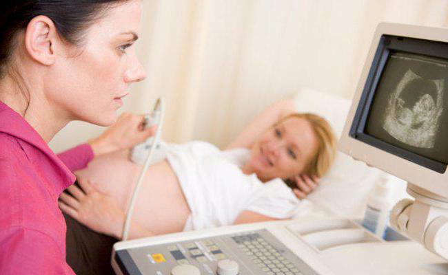
Ultrasound examination is carried out only transabdominal, that is on the skin of the abdomen. To this end, the woman lay down on the couch, opening the stomach. The surface of the belly smeared with gel. The doctor moves the sensor on the front wall. Pain or any discomfort the procedure causes.
And how is biochemical analysis? A pregnant woman comes to the lab on an empty stomach. Blood is taken from a vein in the amount of several milliliters. In the lab the woman provides the results of ultrasound and fills in the questionnaire. The obtained data are processed by the program. The results will only be ready after 14 days.
Transcript
Decoding requires a number of indicators, each of which has its own norm. The result of biochemical screening involves determining the level of these hormones:
- Human chorionic gonadotropin;
- Estriol;
- Fetoprotein;
- Inhibin if a quarter is the triple test.
The Results of the second survey will depend on the duration of pregnancy. They are assessed in accordance with the value of IOM. This is an average figure that is calculated based on the age, body weight of the pregnant woman and the area in which she lives. Indicators diagnostiki divided by the value obtained in the study in a large sample of women of the same age living in the same region.
If the hormone is in the range of 0.5-2.5 Mω, it is considered a normal rate. If the figure is less than this index, or Vice versa above, in this case shown expert advice.
Transcript of diagnosis of the second trimester is the designation of the level of risk in the field of a particular disease. It is designated as a fraction. High risk index of 1:250 or 1:360 for any kind of pathology. So revealed down syndrome, neural tube defects, Edwards, Patau. This requires genetic counseling. With the figure of 1:100 can be offered invasive testing, which involves the detection of suspected pathologies by a set of chromosomes of the embryo.
Some defects can manifest much later and will appear only at the second screening period 16-20 weeks (2nd trimester). Ultrasound in a given period takes into account this feature. In case of negative results of the second screening are advised to seek the advice of a genetics.
Article in other languages:

Alin Trodden - author of the article, editor
"Hi, I'm Alin Trodden. I write texts, read books, and look for impressions. And I'm not bad at telling you about it. I am always happy to participate in interesting projects."
Related News

Snot in dogs: symptoms, treatment and advice of professionals
With the onset of autumn frosts not only the person starts to get sick with colds. Snot the dog – this is not a rare phenomenon, know about it, even novice handlers. However, it can indicate many abnormalities in the body of...

Rickets in puppies: symptoms and treatment
Rickets affects dogs at a young age, when the animals, especially the large breed, rapidly growing. The most dangerous is the age from three months to a year. Pathology may be associated with vitamin D deficiency and newstories ph...

Colds in dogs: symptoms and treatment
Strong the dog's immunity able to fight the virus in the environment. With such protection, the dog can live a long time without fear of catching cold. This disease can often hurt a dog that leads a very active life, and the body ...

Watch Breitling Navitimer: benefits and product features
watches have become a classic item that is a stylish accessory. In the male they are almost the main decoration of confirming the status and character of the owner. Now many firms produce a variety of accessories, which make right...

Leonberger: description of the breed, photo, nature, conditions of detention
the Dog — the dog is large and strong. This loving, sociable, and fearless animal, which is often called «family». The Leonberger is universal: it can be a great companion, alert watchdog, a reliable security gua...

Herpes in cats: possible causes and treatments
unfortunately, no living creature living on our planet, is not immune from acute infections. One of the most common ailments faced by owners of Pets, is a herpes in cats. The symptoms and treatment of this disease will be discusse...
Comments (0)
This article has no comment, be the first!