Today - 18 May 2025
Now - 12:22:12
Now - 12:22:12
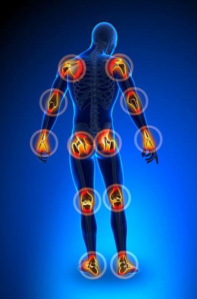
Human bone is so strong that is able to withstand about 10 thousand pounds, but if the skeleton consisted of only one solid bone, our movement would be impossible. Nature has solved this problem by dividing the skeleton into many bones and creating joints-places where bones intersect.
Human Joints perform a quite important function. Thanks to them bones, teeth and cartilage of the body join each other.
They can be classified according to functionality:
Joint which does not allow movement, known as sinetron. The sutures of the skull and gompas (soedinennye teeth with skull) are examples of synarthroses. Connections between bones are called syndesmoses between cartilage synchondroses, bone - centostazioni. Synarthrosis formed with the connective tissue.
Amphiarthrosis allows slight movement of the United bones. Examples of amphiarthroses are the intervertebral disks and pubic symphysis.
The Third functional class – svobodnodispersnye diachrony. They have the highest range of motion. Examples: elbows, knees, shoulders and wrists. It's almost always the synovial joints.
The joints of the human skeleton can also be classified according to their structure (by the material of which they consist):
Fibrous joints are composed of a tough collagen fibers. These include the sutures of the skull and the joint that connects the ulna and radius bones of the forearm together.
The Cartilaginous human joints are composed of cartilage that connect the bones together. Examples of such compounds are the joints between the ribs and costal cartilage, and between intervertebral disks.
Recommended

A tablet from worms – the relevance of the application for the person
How relevant today, drugs against worms in humans? What kind of creatures these worms, what are modern methods of treatment? We will try to answer these questions, since ignorance in this area is undesirable. Imagine a mummy, which is misleading in k...

What to do if you cracked skin on hands?
Each of us at least once in a lifetime encounter with a small, but very, when the crack the skin on the hands. At this time there are wounds of different sizes, which hurt and cause inconvenience, especially when in contact with water or detergents. ...

Spray Macho man - the key to a proper relationship between the two spouses
Male impotence is a pathological condition associated with abnormal physiological capacity of the penis to reginout and bring sexual partner pleasure in bed.sex impotenceimpotence may not men to pass unnoticed – it usually spoils his nervous sy...
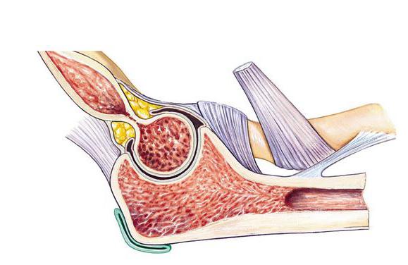
The Most common type of synovial joint is a fluid-filled space between the ends of the associated bones. It is surrounded by a tough capsule of dense connective tissue covered with synovial membrane. The synovial membrane, which consists of the capsule, produces synovial fluid the oil, the function of which is to lubricate the joint, reducing friction and wear.
There are several classes of synovial joints, for example, ellipsoidal, ginglymoid, saddle-shaped, and spherical.
Ellipsoidal joints connect bones and smooth allowing them to slide past each other in any direction.
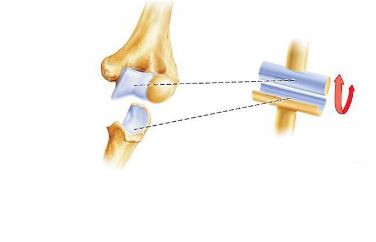
Hinge joints, e.g. elbow and knee joint of a person, restrict movement in only one direction so that the angle between the bones is possible to increase or decrease. Restricted movement in the hinge joints provides for more strength and toughness to the bones, muscles, and ligaments.
Saddle joints, such as between the first metacarpal bone and the bone, trapezium, bones allow you to turn 360 degrees.
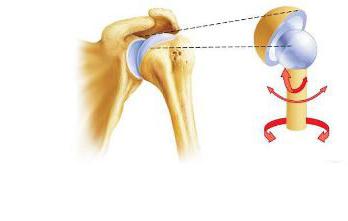
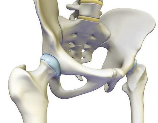
Shoulder and hip joint in the human body – the only spherical joints in the body. They have the free range of motion, they are the only ones that can be rotated around its axis. However, the lack of spherical joints is that free range of movement makes them more susceptible to dislocation than the less movable joints of a person. These places are often fractures.
Some synovial types of human joints must be considered separately.
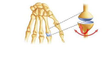
Ginglymoid joints are synovial class. It is ankle, knee and elbow joint of the person. Usually a hinge joint linking two or more bones where they can only move in one axis to bend or straighten up.
The simplest hinge joints in the body - interphalangeal, they are located between the phalanges of the hands and feet.
Because they have little body weight and mechanical force, they consist of a simple synovial material with a tiny additional ligaments to strengthen. Each bone is covered with a thin layer of smooth hyaline cartilage, is designed to reduce friction in the joints. Bone is also surrounded by a capsule of tough fibrous connective tissue covered with synovial membrane.
The structure of the joint person is always different. For example, the elbow joint is more complex, is formed between the humerus, radius and ulna bones of the forearm. The elbow is subjected to stronger stresses than joints in the fingers and toes, therefore contains several additional ligaments strong and unique bone structures that reinforce its structure.
The Ulnar and radial collateral ligaments help to hold the ulna and radius bones and strengthen the joints. The legs of a man also consist of several large hinge joint.
Like elbow ankle joint is located between the large and small tibial bones in the lower leg and the talus in the foot. Branches tibia fibula bones form the bony socket around the talus to limit the foot traffic in one axis. Four additional ligaments, including the deltoid, fasten the bone and strengthen the joint to support the weight of the body.
Located between the thigh and the leg between the tibia and fibula bone of the lower leg, the knee joint is the largest and most complicated hinge joint in the human body.
Elbow joint and ankle joint, the anatomy of which are similar, are most often susceptible to osteoarthritis.
Ellipsoid joint, alsoknown as flat, is the most common form synovial joints. They are formed near the bone with smooth or almost smooth surface. These joints allow the bones to glide in any direction - up and down, left and right, diagonally.
Due to its structure ellipsoidal joints flexible, and their movement is limited (to prevent injury). Ellipsoidal joints sinovialnoj covered with a membrane that produces fluid, which serves as a lubricant for the joint.
Most ellipsoidal joints are found in appendicular skeleton between the carpal bones of the wrist, between the carpal joint and the metacarpal bones of the brush, between the bones of the ankle.
Another group of ellipsoidal joint is located between the faces of twenty-six vertebrae in the intervertebral joints. These joints allow us to bend, straighten and rotate your torso, while keeping the strength of the spine which supports the body weight and protects the spinal cord.
There is a separate variety of the ellipsoid joints - condyloid joint. It can be considered a form of transition from a hinge type of joint to the ellipsoid. From ginglymoid condylar joint is characterized by great difference in the shape and dimensions of the mating surfaces, resulting in possible movement around two axes. From ellipsoidal joint the condylar differs only in the number of joint heads.
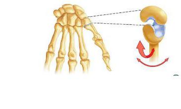
Saddle joint is a type of synovial joints, where one bone shaped like a saddle and the other bone relies on it like a rider on a horse.
Saddle joints are more flexible than spherical or ellipsoid.
The Best example of a saddle joint in the body is the carpometacarpal joint of the thumb that is formed between the trapezium bone and the first metacarpal bone. In this example, the trapezium forms a rounded saddle, which sits on the first metacarpal bone. Carpometacarpal joint allows the thumb easy to promote with the other four fingers of the hand. Thumb, of course, extremely important to us as it allows our hand to firmly grasp objects and use many tools.
Ball joints are a special class of synovial joints that have the highest freedom of motion in the body thanks to their unique structure. Hip joint and shoulder joint are the only globular in the human body.
The Two main components of spherical joint, a bone with a spherical head and a bone with a bowl-shaped recess. Consider the shoulder joint. Human anatomy is so arranged that the spherical head of the humerus (upper arm bone) fits into the articular cavity of the scapula. The glenoid cavity is small and shallow recess, through which the shoulder joint the greatest range of motion in the human body. It is surrounded by a ring of hyaline cartilage that are flexible to strengthen the bones, while muscles - rotator cuff - hold the humerus within the socket.
The Hip joint is somewhat less mobile than the shoulder, but is stronger and more stable joint. Additional stability to the hip joint required to withstand the weight of the human body on the feet, performing actions such as walking, running, etc.
In the hip joint rounded, almost spherical head of the femur (thigh bone) fits snugly to the acetabulum, the deep cavity in the pelvic bone. A sufficiently large number of strong ligaments and strong muscles keep the head of the femur in place and resist the strong stresses in the body. Acetabulum also prevents the dislocations of the thigh, restricting the movement of the bone within it.
Based On the foregoing we can make a small table. The structure of the joint in the human body to include in it will not. So, in the first column of the table indicates the type of the joint, the second and the third – examples and their location, respectively.
Type of joint | Examples of joints | Where |
Ginglymoid | The Knee, elbow, ankle. The anatomy of some of them below. | Knee – between the femoral, tibial bones and patella; ulnar – between the humerus, ulna and radius bone; the ankle-between leg and foot. |
Ellipsoidal | The Intervertebral joints; the joints between the phalanges. | Between the faces of the vertebrae; between the phalanges of feet and hands. |
Globular | Hip and shoulder joint. Human anatomy pays this kind of joints special attention. | Between the femur and pelvic bone between the humerus and scapula. |
Saddle | The Carpal-metacarpal. | A bone Between the trapezium and the first metacarpalbone. |
To make it clearer what constitutes a human joint will describe some of them.
The Elbow joints human anatomy which have already been mentioned, require special attention.
The Elbow joint is one of the most complex joints of the human body. It is formed between the distal end of the humerus (more precisely, its articular surfaces - block and the condyles), the radius and the trochlear cut out the ulna, and the head of the radius and its articular circumference. It consists from three joints: the brachioradialis, preselective and the proximal radioulnar.
Preselective joint is located between the trochlear notch of the ulna and the unit (articular surface) of the humerus. This refers to a hinge joint is uniaxial.
The Brachioradialis joint is formed between the condyles of the humerus and humeral head. Movement in the joint occur around two axes.
Premaxillary radioulnar connects the radial notch of the ulna and the articular circumference of the head of the radius. He also uniaxial.
At the elbow joint, no lateral movement. In General, it is considered a hinge joint with a helical shape of the slide.
The largest of the upper body are considered to be the elbow joints. The legs of a man is also composed of joints about which simply can not tell.
This joint is located between the acetabular cavity of the pelvis and the femur (her head).
This head is covered with hyaline cartilage throughout, except for the holes. Acetabulum is also covered with cartilage, but only about the lunate surface, the remaining part is covered sinovialnoj membrane.
For the hip joint are the following ligaments: the sciatic-femoral, ilio-femoral, pubic-femoral, circular area, and the ligament of the femoral head.
Ilio-femoral ligament originates on the lower anterior iliac spine and ends at the intertrochanteric line. This bunch is involved in maintaining the torso in an upright position.
The Next ligament, sciatic-femoral, begins at the ischium and is woven into the capsule of the hip joint.
Just above the top of the pubic bone, starts pubic-femoral ligament, which runs down to the capsule of the hip joint.
Within the joint is the ligament of the femoral head. It takes from the beginning of the transverse ligament of the acetabulum and ends at the fovea of the femoral head.
The Circular area is made in the form of a loop: it is attached to the lower front iliac bone and surrounds the neck of the femur.
Hip and shoulder joints are the only globular in the human body.
This joint consists of three bones: the patella, the distal end of the femur and the proximal end of the tibia.
The Capsule of the knee joint attached to the edges of the tibia, femoral bone and patella. To the femur, it is secured under namesake. On the tibia is fixed at the edge of the articular surface and patella the capsule is attached in such a way that all its front surface is out of joint.
The ligaments of the joint can be divided into two groups: intracapsular and uncapable. Also in the joint there are two lateral-tibial and fibular collateral ligament.
It is formed with the articular surface of the talus and the articular surfaces of the distal ends of the fibula and tibia.
The Articular capsule almost all along attached to the edge of the articular cartilage and moves away from him only on the anterior surface of the talus. On the lateral surfaces of the joint are ligament.
The Deltoid or medial ligament, consists of several parts:
- posterior tibial-talus, between the posterior margin of the medial malleolus and posterior medial divisions of the talus;
- anterior tibial-talus, situated between the anterior edge of the medial malleolus and the posteromedial surface of the talus;
- tibial-heel portion extends from the medial malleolus to the talus supports;
- tibio-navicular part, originates from the medial malleolus and ends at the rear surface of the navicular bone.
The Next ligament, calcaneal-fibular, extends from the outer surface of the lateral malleolus to the lateral surface of the neck of the talus.
Close to the previous one is the anterior talofibular ligament between the front edge of the lateral malleolus and lateral surface of the neck of the talus.
Finally, the posterior talofibular ligament originates at the posterior edge of the lateral malleolus and ends at the tubercle of the lateral process of the talus.
In General, the ankle joint is an example of a hinge joint with a helical movement.
So now we have an idea of what human joints. Anatomy of the joints is harder than it looks, and you yourself can verify this.
Article in other languages:
AR: https://tostpost.com/ar/health/17487-the-human-joints-types-of-human-joints.html
BE: https://tostpost.com/be/zdaro-e/33672-sustavy-chalaveka-v-dy-sustava-chalaveka.html
HI: https://tostpost.com/hi/health/19127-the-human-joints-types-of-human-joints.html
JA: https://tostpost.com/ja/health/17145-the-human-joints-types-of-human-joints.html
KK: https://tostpost.com/kk/densauly/33985-buyndar-adam-t-rler-buyndardy-adam.html
PL: https://tostpost.com/pl/zdrowie/35034-stawy-cz-owieka-rodzaje-staw-w-cz-owieka.html
PT: https://tostpost.com/pt/sa-de/34832-as-articula-es-do-homem-tipos-de-articula-es-pessoa.html
TR: https://tostpost.com/tr/sa-l-k/30402-eklemler-insan-t-rl-eklem-ki-i.html
UK: https://tostpost.com/uk/zdorov-ya/34197-suglobi-lyudini-vidi-suglob-v-lyudini.html
ZH: https://tostpost.com/zh/health/7273-the-human-joints-types-of-human-joints.html

Alin Trodden - author of the article, editor
"Hi, I'm Alin Trodden. I write texts, read books, and look for impressions. And I'm not bad at telling you about it. I am always happy to participate in interesting projects."
Related News

Proper oral hygiene for a dazzling smile
a Dazzling, white smile in the first place, a measure of health. And our relationship to him. To take care of their teeth, we start from the childhood. But the older a person is, the more care is required by the body. And if in th...

Hookah – it's art embodied the Eastern tradition, won the whole world. For many centuries Eastern sheiks smoked it around. The first such device was a coconut, but gradually the art of making Shisha has reached incredible he...

Set on edge – what is this? The meaning of the word "mouth"
This word was formed from an obsolete verb “skomal”, which was used in the past. It meant “to ache, hurt”. Alternated with the verb “sting”. Gradually the meaning of some words have changed, som...

What administered injections osteochondrosis of the cervical spine? Recommendations and treatment
Statistics show that diseases caused by premature aging, wear and tear of tissues, today not only affect the elderly. Increasingly for help to doctors, patients young and middle age. Pathology of musculoskeletal system considerabl...

Why do women need to exercise the muscles of the vagina
the Benefits of sports are obvious to everyone: good health plus beautiful body. Many women are willing to spend hours to visit a gym, hairdresser and shops. Appearance is, of course, wonderful and attracts the attention of men, b...

"Fenoterol": instructions for use, contraindications and procedure of admission of
asthma, lung disease, hypertonicity of the uterus - the unifying feature of these pathologies is the increased muscle tension. To resolve this condition, use different medication. This remedy is the "Fenoterol". Application instru...


Comments (0)
This article has no comment, be the first!