


The x-rays: description of procedure, transcript and recommendations
Table of contents:
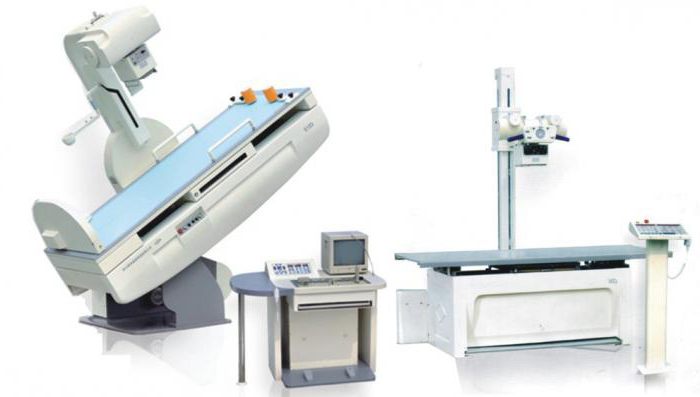
Radiography is one of the methods of research, the basis of it – obtaining a fixed image by means of x-rays. Usually get the result on x-ray film or withdrawn (if used for digital devices) on the screen or paper. The study is based on passing x-rays through the tissues of the body. Typically use x-rays as a diagnostic method. For more accurate results use of x-rays in two projections.
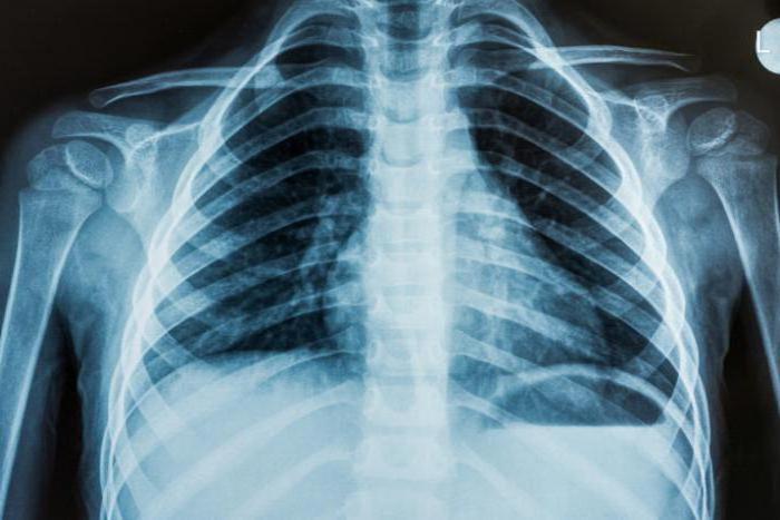
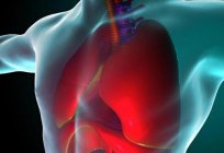
Chest x-ray
Radiography OGK (chest) – the most common method of survey that allows you to identify pathology of the respiratory and cardiovascular systems, ribs, thoracic spine, occurring in various injuries and diseases.
How are x-rays? Passing through the body and organs they are absorbed in different ways. The result is the x-ray. Tissue more dense structures appear white on it, those are softer – dark. After development and drying, the radiologist evaluates the resulting picture. X-ray picture of the lungs will show all pathology, if any, will indicate disease.
Modern digital cameras simplify the procedure, the radiation dose is significantly reduced. And there is a mobile equipment that allows you to examine bedridden patients.
Possible x-rays and decoding of the result
Chest x-ray helps detect the following pathologies in the body:
- Respiratory system: bronchitis, pulmonary fibrosis, pleurisy, tuberculosis, cancer, lung atelectasis, pneumonia. X-rays decrypts the doctor can immediately see the likely disease.
- Cardiovascular system: myocarditis, pericarditis, heart changes in size.
- The Mediastinum: displacement structures, mediastinitis.
- The musculoskeletal skeleton of the thorax: fractures of the sternum or ribs, vertebrae, hemothorax, pneumothorax, injury of the mediastinum and heart.
Radiography is used for tracking the dynamics of recovery in the treatment of pneumonia. However x-rays cannot be called a universal method of diagnostics. For example, the nature of the tumor x-rays to assess you can't, also this study is limited to stationary patients. For such exceptional cases, computed tomography is used.
When the decoding result x-ray picture OGC doctor evaluates what the size and shape of the mediastinum, the structure of the thorax and soft tissues, transparency of the lung fields, the intensity of the figure, the position and structure of roots of the lungs, form of the pleural sinus and diaphragmatic domes.
Preparation and holding procedure
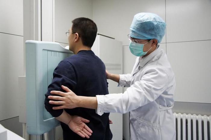
For the procedure radiography OGK does not require special training. The doctor recommends to remove clothing and jewelry from the area that is to be irradiated. You also need to remove all items that may interfere with the study (eyeglasses, dentures). If there is a need for the presence of a relative of the patient, it is worn on a protective lead apron.
Removing his clothes, the patient is positioned in front of the photographic plates. The doctor leaves the room to the console, his team needed to lift their shoulders, cuddling up to the plate and hold your breath for a while. To move this is not. If the patient cannot take a vertical position, it is placed on the table. Help him in this case, the relatives or the nurse.
The Examination is painless, does not cause any discomfort. The only discomfort – a cool room temperature. The x-rays will be ready within 15 minutes. It will give you immediately along with a description. Based on this, the doctor will make a diagnosis or refer for further examination.
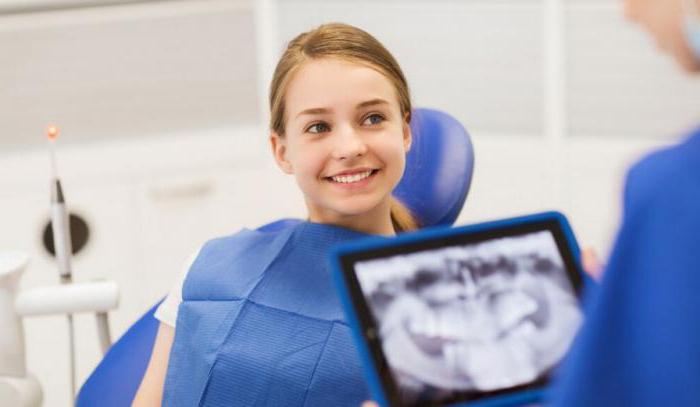
X-ray pictures of teeth
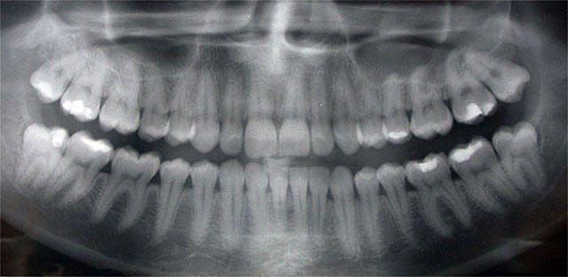
X-ray examination is widespread in dentistry. The not only provides the ability to track pathology, but also reveals deviations in the structure of the jaw. X-ray diagnosis is important in selecting optimal treatment options.
There are several types of x-ray snapshot in dentistry
- Panoramic. This image allows the doctor to evaluate the panorama of the entire dentition, to determine their number, to see the presence of impacted teeth, the beginnings. Also shows the anatomical structure of the jaw, sinus. A panoramic image is important in implantation of teeth, bite correction, removal of wisdom teeth.
- Bite. Otherwisethis is called the interdental radiography. Common type of the image. It is used to identify periodontitis, dental caries. Sometimes a bite is done after the installation of crowns to verify correct procedure.
- Aimed. With the sighting of the image we can see exactly how does a bad tooth, establish the correct treatment regimen. X-ray allows you to see not more than four teeth.
- Digital. Safe and modern diagnostics. 3D-imaging enables to obtain a clear picture of the entire dentition and individual teeth. A three-dimensional image is displayed on the screen after the study doctor determines treatment methods.
Procedure of execution of the picture
X-ray scan of the teeth is performed according to the recommendations of the dentist: in case of detection of caries, abnormal occlusion, diseases of periodontal tissues, with pulpitis, cyst, injuries of the jaw, abscesses.
The study recommended the patient to remove all metal items, jewelry: they can distort the pictures. The procedure depends on the type of the image. Is the study a few minutes. The radiation is minimal dose. The session takes place in a special room. The patient occludes the photosensitive film, it must be between the machine and examined the tooth.
In the study with computer radiovisiograph the patient must put on a special apron, the sensor is installed on the investigated area and is attached to the apparatus. The result is displayed on the computer.
The use of orthopantomograph radiograph is performed in the following way: the patient is to the camera, the chin is fixed on the support. Teeth clamped unit which does not close the jaws. The patient must be stationary. The device rotates several times around the head. Images can be taken on the same day.
Transcript of a snapshot
On the basis of x-ray picture of teeth, the doctor writes the conclusion, which indicates the number of teeth, size and their location. All detected abnormalities will also appear in the conclusion.
The picture shows the location of each tooth, the tilting condition of the bones. Dimming in the picture indicate the presence of pulpitis, denticle. Defects in tooth enamel indicate tooth decay. Where the density is reduced, the visible illumination. If the tooth is difficult, the tooth structure is deformed, the formation of granulomas.
Can be discovered a cyst – a clear outline of the uniform structure of elongated shape. Cysts located at the tooth root, it can be small and large. Large cysts can affect just two teeth. Chronic periodontitis is seen as a darkening in the picture at the top of the root. When periodontal disease is reduced bone marrow visible region, visible atrophic and sclerotic processes of change.
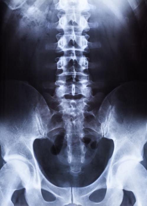
X-ray of spine
In some cases, the doctor recommends to do the x-rays of the spine?
- When pain in the cervical, thoracic and lumbar.
- Lumbar muscular pain of obscure nature.
- By limiting the mobility of the limbs.
- Injuries, falls and bruises.
- If you suspect degenerative changes in the bones.
- In the diagnosis of curvature, osteochondrosis, scoliosis.
The x-rays it is recommended to perform two projections: lateral and frontal. Description x-ray image makes the radiologist, he estimates the contours of the vertebrae, the spaces between them, the intensity of the color, the presence of growths. After that an experienced specialist are able to diagnose, to determine the probable prognosis and the need for surgical treatment.
How the procedure
For a picture of the upper spine does not require special training. If we study the lumbar-sacral region, it is recommended to prepare in advance:
- You Need to completely clean the intestines, otherwise the diagnosis will be difficult to put right.
- To Exclude from the diet for two days prior to the procedure products that contribute to fermentation: bread, milk, pulses, coarse fiber.
- Should be deleted Before dinner, before the procedure – Breakfast.
- To Give up alcohol and Smoking.
- Before the procedure to cleanse the bowel with an enema.
- At the time of shooting on the body should not be metallic objects.
- Keep a steady position.
The Examination for the patient is absolutely painless. Held over 10-15 minutes. Pictures with description immediately handed out.
Article in other languages:
PL: https://tostpost.com/pl/zdrowie/22717-rtg-zdj-cie-opis-procedury-definicje-i-zalecenia.html
TR: https://tostpost.com/tr/sa-l-k/22767-x-ray-resim-a-klama-bak-mlar-de-ifre-ve-neriler.html
ZH: https://tostpost.com/zh/health/13459-the-x-ray-procedure-explanation-and-recommendations.html

Alin Trodden - author of the article, editor
"Hi, I'm Alin Trodden. I write texts, read books, and look for impressions. And I'm not bad at telling you about it. I am always happy to participate in interesting projects."
Related News

Monarda oil: properties and applications
For many people, the aromatic oils are integral part of life. Their impact on health and General wellbeing proven for a long time. Some esters are used in everyday life, the other is used for body care or for medicinal purposes, b...

We will figure out why your period came early
There are many answers to the question about why your period came early. I'll try to list them.the First and most common cause of earlier onset of period bleeding is irregular menstrual cycle. Needless to say, that those girls who...
When there is a rash in syphilis? If itchy rash in syphilis?
One of the most unpleasant symptoms of infectious diseases is a rash in syphilis (photo vividly captures the essence of the problem). Such education can significantly spoil the appearance of the patient and will even go as ulcers....
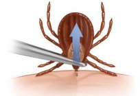
How to remove a tick with yourself?
People who have suffered from the bites of these parasites are often asked about how to remove a tick per person, and you can do it yourself? To not simmer long wait, just answer this question: of course you can. Let's try to unde...

Pituitary adenoma. Symptoms, classification
pituitary Adenoma is a tumor of benign nature. The tumor originates from cells in the anterior pituitary lobe. The tumor is localized in the region of Turkish saddle in the skull base (sphenoid part of it). Typically, a pituitary ...

The drug "Pentas": instructions for use, analogs, reviews and
dose of prescribed medication “the Pentas”? Instruction manual, reviews and the drug is considered next. Also the article presents information on the release of this medication and its properties.Structure, forms of me...
Comments (0)
This article has no comment, be the first!