


Visual evoked potentials. The vision test on computer
Table of contents:
Visual evoked potentials – potential biological nature that appear in the cortex as a response to the effect of light on the retina.
A Little history
They were First described by E. D. Adrian in 1941, however, steadily fixed them after Davis and Galambos advanced method of summation of potentials in 1943. Then the registration method ZVP has found wide application in the clinic where he explored the functional position of the visual pathway in patients ofthalmoplegical sphere. To register ZVP, used standard electrophysiological specialized systems based on modern computers.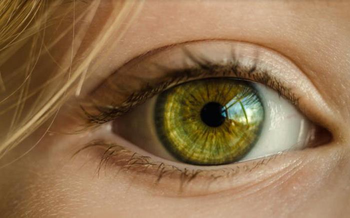
The plaque, i.e., active electrode placed on the patient's head an inch above the tuberosity of the occiput in the midline over the area where the visual striate cortex is projected to the cranial vault. The second indifferent electrode mounted on the ear lobe or mastoid. On the other lobe of the ear or on the skin in the middle of the forehead fasten the grounding electrode. How is eyesight test on computer? As a stimulant is used or flash light (flare PEL), or reversible patterns on the monitor (pattern-VEP). Stimulating the field of view has a size of approximately fifteen degrees. Research is carried out without an increase in pupils. The role also and the age of the person subjected to the procedure. Look at how he sees people.
Learn More about the concept
ZVP are bioelectric response of the visual areas in the cerebral cortex and thalamocortical pathways and subcortical nuclei. Wave generation ZVP has a connection also with the generalized mechanisms of brain activity spontaneous character which is recorded on the EEG. Responding to the effects of light on the eye, vecs show bioelectric activity mainly in the macular areas of the retina, due to its large representation in visual cortical centres in comparison with the regions of the retina located at the periphery.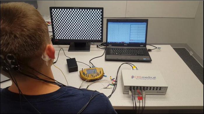
Recommended

A tablet from worms – the relevance of the application for the person
How relevant today, drugs against worms in humans? What kind of creatures these worms, what are modern methods of treatment? We will try to answer these questions, since ignorance in this area is undesirable. Imagine a mummy, which is misleading in k...

What to do if you cracked skin on hands?
Each of us at least once in a lifetime encounter with a small, but very, when the crack the skin on the hands. At this time there are wounds of different sizes, which hurt and cause inconvenience, especially when in contact with water or detergents. ...

Spray Macho man - the key to a proper relationship between the two spouses
Male impotence is a pathological condition associated with abnormal physiological capacity of the penis to reginout and bring sexual partner pleasure in bed.sex impotenceimpotence may not men to pass unnoticed – it usually spoils his nervous sy...
How does registration work?
Check the visual evoked potentials carried out in the form of electric potential fluctuations consistent or components that differ in polarity: negative potential, or the N is oriented upwards, the positive potential, that is, P down. Characteristic of VEP contains a shape and two numbers. Potentials VEP largest in the norm is much less (approximately 40 mV) in comparison with the waves of the electroencephalogram (up to 100 µv). The definition of latency is the time period from the start of the light stimulus until the maximum performance potential of the cortex. Most often, the potential reaches its maximum value after 100 MS. If there are different kinds of pathology of the visual pathway, the VEP shape changes, the amplitude of the components decreases, the latency is lengthened, that is, increasing the time during which the pulse passes to the cortex of the brain through the optic path.
In what lobe is the visual area? It is located in the occipital lobe of the brain.
Varieties
The nature of the components in the VEP and their sequence is fairly stable, however, the temporal characteristics and amplitude of normal have variations. This is determined by the conditions in which to conduct the study, the specifics of the light stimulus, an overlap of the electrodes. During stimulation of semi-fields of view and reverse frequency one to four times per second for registration of phasic transient-VEP, which has consistently highlighted three components – N 70, R 100 and N 150. The frequency of reversion by increasing more than four times per second causes the rhythmic of the total response in the cortex in the form of a sine wave that ZVP is called the stability condition in steady-state. These potentials differ from the phasic to the fact that they are no sequential components. They look like a rhythmic curve with alternating reducing and UPS capacity.
Normal values visual evoked potential
ZVP Analysis is carried out according to the amplitude of the potentials measured in micro volts, the form of entry and the time period from exposure to light prior to the occurrence of the peaks of the waves ZVM (calculated in milliseconds). Also pay attention to the difference between the amplitude potential and the value of the latency of light stimulation in the right and left eye alternately.
In ZVP (which is in ophthalmology, it is interesting to many) a phasic-type during reversion from the low frequency checkerboard pattern or in response to a light flash with special constancy allocated R 100, a positive component. The duration of the latent period of this component varies the rate of from ninety-five to one hundred and twenty milliseconds (cortical). The preceding component, i.e., N 70, - from sixty to eighty milliseconds, N 150 – from one hundred and fifty to two hundred. Late R 200 are not recorded in all cases. So passes the eye test on the computer.
Since the amplitude of the VEP is characterized by its variability, taking into account the results of the study she has a relative value. Normal values for its value in relation to R 100vary for an adult, from fifteen to twenty-five microvolts, a higher potential value in children – up to forty microvolts. For pattern stimulation, the amplitude value of ZVP is a little lower and is determined by the size of the pattern. If the amount of squares more, the potential is higher and Vice versa.
Thus, visual evoked potentials are a reflection of the functional state of ways to view and provide information of a quantitative nature in the course of the study. The results allow us to diagnose the pathology of the visual pathways in patients neuro-ophthalmology field.
Here's how to see people.
Topographic mapping of brain potentials in the head-ZVP
Topographic mapping of brain potentials in the head of multichannel VEP records the potentials from different brain regions: parietal, frontal, temporal and occipital. The results of the study are transmitted to the monitor screen as topographic maps in colour which varies from red to blue. Topographic mapping shows the amplitude value of the potential of ZVP in ophthalmology. That is, we explained.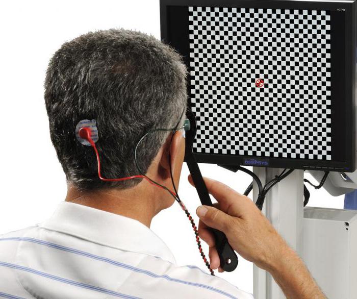
The head of the patient must wear special helmet with sixteen electrodes (the same as for EEG). Installing electrodes on the scalp at particular points in the projection: the parietal, frontal, above the left and right hemispheres, temporal and occipital. Processing and recording of biopotentials is carried out using electrophysiological specialized systems, for example, “Neurocirculate” the company "MBN". Through this technique allows the electrophysiological differential diagnosis in patients. During retrobulbar neuritis acute forms, on the contrary, it is noted bioelectrical activity, receiving expression in the neck, and the almost complete absence of excited sites in the frontal lobe of the brain.
Diagnostic value of visual evoked potentials in different disease
In physiological and clinical studies, if visual acuity is quite high, it is best to apply the method of registration of individuals ZVP on reversion.
In clinical and physiological studies in high visual acuity, it is preferable to use a method of registration of individuals VEP to reversing checkerboard patterns. These potentials are quite stable in terms of amplitude and temporal properties are well reproducible and are sensitive to various abnormalities in the visual pathways.
On the same flash VEP are characterized by more variability and lower sensitivity to change. This method is used when a serious decline in visual acuity of the patient, the lack of bracing of a view, with an impressive clouding of the eye optical media, nystagmus pronounced character, and in young children.
In the test for vision screening involved the following criteria:
- Lack of response or a large decrease in amplitude;
- The longer the latency of all the highlights of the potentials.
Recording visual evoked potentials, it is necessary to take into account the normal age, especially for study of children. Interpreting information registration ZVP in early childhood with abnormalities of the visual pathways, we should consider the characteristics of electrocortical reaction.
There are two separate phases in the development of ZVP, which are recorded responsively to the pattern reversion:
- Quick – from birth to six months;
- Slow & ndash; from six months until puberty.
In the first days of life children are registered ZVP.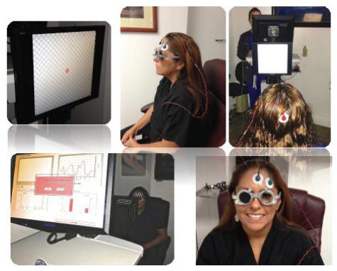
Topical diagnostics of pathology of brain
That shows EEG? Chiasm at the level of pathology of the visual pathways (tumor, trauma, optiganally arachnoiditis, demyelinating processes of the aneurysm) there was a decrease in the amplitude of potentials, the latency increases, the individual elements of ZVP fall. Increasing changes in the VEP simultaneously with progression of the lesion. In the pathological process involved precisely district of the optic nerve, as evidenced by ophthalmoscopically.
Retronasally pathology differ interhemispheric asymmetry of the visual capabilities and are better seen for multichannel record type topograficheskiy mapping.
Chiasm lesion is characterized by an asymmetry of VEP crossed character, reflected in the significant changes of the biopotentials in the brain on the opposite side from the eye with reduced visual functions.
During the analysis of the SGP should take into account hemianopsia loss of the visual field. In this connection, when chiasm pathologies stimulation half of the visual field increases the sensitivity of the method, allowing to reveal the distinctive features between the dysfunction in the fibers of view, that go from the nasal and temporal parts of both retinas of the eye.
Retropatellar level of defects of the visual pathways (bundle Grazioli, the optic tract, the visual area of the cerebral cortex of the head) observed dysfunction of the unilateral nature, manifested in the form supercriminal asymmetry, which is expressed in pathological VEP with the same indicators in the stimulation of each eye.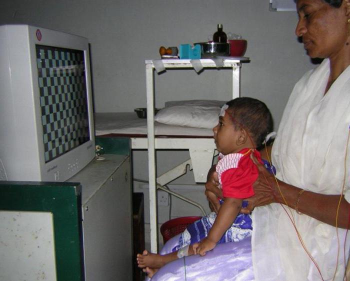
The Reason is reduced bioelectrical activity of neurons in Central visual pathways - homonymous defects of the visual field. If they produce the capture of the macular region, while stimulating the half of the field changed and get the form which is characteristic of the Central cattle. If the primary visual centers continue, ZVP can have normal levels. Still that shows EEG?
Pathology of the optic nerve
If in the optic nerve are the pathological processes, the most characteristic of their manifestation is the increase of the latency of the main component of VEP P 100.
Optic Neuritis from the affected eye along with the increase of latency is characterized by a decrease of the amplitude of the potentials and the change in components. That is, the Central vision is impaired.
Often check the W-shaped component R 100 associated with a decrease in functioning of the axial bundle of fibers of nerves in part of the optic nerve. The disease progresses along with the increase in time latency of thirty to thirty-five percent, decrease in amplitude and formal changes in the components of VEP. If the inflammatory process subsides in the optic nerve, and visual function increased, the shape of ZVP and amplitude values are normalized. The temporal characteristics of ZVP remain enlarged for two to three years.
Optic Neuritis, which develops on the background of multiple sclerosis, is determined to identify clinical symptoms according to the changes in VEP, indicating early involvement of visual pathways in the pathological process.
The Defeat of the optic nerve unilateral nature, it has very significant differences in the latency component R 100 (twenty-one millisecond).
Front and rear ischaemia of the optic nerve because of the acute defect of arterial circulation in the vessels which nourish it, are accompanied with the patient's eyes a noticeable decrease in the amplitude of VEP and not too tall (three milliseconds), the increase in the latency of P 100. The indicators ZVP healthy eyes usually remain normal.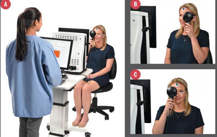
Stagnant disk at the initial stage is characterized by reduction in amplitude of visual evoked potentials (VEP) of a temperate character and a small increase in latency. If the disease progresses, some violations receive an even more tangible expression that is fully consistent with the picture of Ophthalmoscope.
When atrophy of the optic nerve secondary type after the transfer of ischemia, neuritis, congestive disk and other pathological processes are also observed decrease of indicators of VEP amplitude and an extension of time of latency P 100. Such changes can be characterized by a different degree of expression and to appear independently from each other.
Pathological processes in the retina and choroidal (Central serous homeopatia, the multiple forms of maculopathy, macular degeneration) increases the latency period and decrease in the amplitude of the potentials.
Often not observed correlation between the decrease in amplitude and increase in length of latency potentials.
Conclusion
So, we can conclude that although the method of analysis of the VEP is not a specific in the determination of any pathological process visual path, it is used for early diagnosis in the clinic of various diseases of the eye and clarify the extent and level of injury. Of particular importance is a test to check your vision and in ophthalmic surgery.
Article in other languages:
AR: https://tostpost.com/ar/health/4314-visual-evoked-potentials-the-vision-test-on-computer.html
DE: https://tostpost.com/de/gesundheit/7651-evozierte-visuelle-potentiale-sehtest-am-computer.html
HI: https://tostpost.com/hi/health/4320-visual-evoked-potentials-the-vision-test-on-computer.html
JA: https://tostpost.com/ja/health/4315-visual-evoked-potentials-the-vision-test-on-computer.html
KK: https://tostpost.com/kk/densauly/7653-tuynda-an-k-ru-leuet-tekseru-k-ru-komp-yuterde.html

Alin Trodden - author of the article, editor
"Hi, I'm Alin Trodden. I write texts, read books, and look for impressions. And I'm not bad at telling you about it. I am always happy to participate in interesting projects."
Related News

Allergic to the patch: symptoms and treatment
today, Allergy is one of the most common human diseases. Of course, medicine does not stand still, every day progress is moving forward, but to this point treatment and even detection of allergic reactions at first remains an open...

Pearl barley. Useful properties and application in cooking
All known pearl barley is a delicious grain. For many people it is associated with bread, soups, cereals, and beer. But besides all this, it is also used when you create a different very tasty salads. Barley goes well with spicy s...

it's No secret that the body of any living creature, including man, on 70% consists mainly of water. This is what, amazing, liquid substance that has no color, odor plays a major role in the life of the organism. Without food you ...

Glucose tolerance - what is it?
Problems with the endocrine system lead to the fact that failures occur in all internal organs. As a result, to find the cause, the doctor will assign a number of studies. Among the masses of a great variety of tests will also be ...

Diagnosis of toxoplasmosis. PCR analysis (toxoplasmosis): results and transcript
Scientists say that seventy percent of people on our planet are infected with parasites, the most common of which is the Toxoplasma gondii (Toxoplasma gondii). Many people have probably heard scary stories about the disease. But i...
Telangiectasia or spider veins – the so-called clusters of small veins dark, such as red or purple in color. They may appear on different parts of the body, including the face. To many the question arises whether it is possi...
Comments (0)
This article has no comment, be the first!